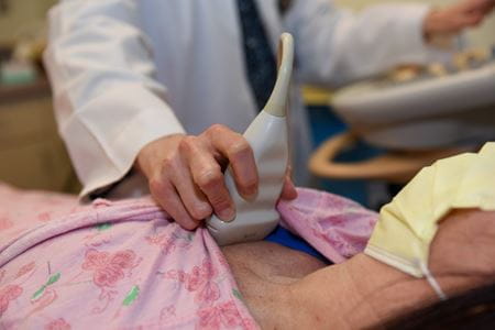
What is ultrasound?
Sometimes referred to as sonography, ultrasound sends sound waves which then bounce off objects within the body and relay back to the sonographer the size of the object and whether it’s moving. For centuries, this technology has also been witnessed in nature where animals use sonar to send sound waves and interpret the returning echoes to detect distance and possible movements in objects. Since its application within medicine, the use of ultrasound has enabled medical experts the ability to capture a moment in time for patients, effectively visualizing anatomy and pathology otherwise unseen without a scalpel.
While ultrasound is commonly known for its use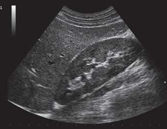 during pregnancy, it plays a vital role in countless other applications such as vascular, abdominal and cardiac imaging. Ultrasound guided procedures allow clinicians to properly identify blood vessels before inserting intravenous catheters, to correctly guide needles during a biopsy or to see heart structures in motion before doing more extensive cardiac procedures.
In the past 20 years alone, ultrasound technology has advanced from being a washing machine-sized device to being something small enough to fit in someone’s hand. The price has gone down and the quality has improved exponentially, enabling experts to more easily bring this technology to more patients. It’s even possible that ultrasound technology may someday be as essential to medicine as the stethoscope.
Education that stands out among the rest
Currently, IU School of Medicine is leading the charge in the advancements in ultrasound technology and its training. The Emergency Ultrasound and the Pediatric Emergency Ultrasound Fellowship Programs not only provide hands-on training for fellows, but they continue to be ranked as some of the best programs in the country offering this specialty. Additionally, the school’s point of care ultrasound (PoCUS) program has helped instill the significant role ultrasound plays in medicine by preparing medical students with the training they’ll need as they continue into residency and clinical settings.
Where IU School of Medicine stands out among the rest is that it also supports the community need for more highly trained sonographers. Currently, the school has an undergraduate Diagnostic Sonography Program at its Fort Wayne campus and a new Diagnostic Sonography Program at its Indianapolis campus beginning in 2023. With the advancements in technology and the increasing need for expertise in the area of ultrasound, Peterson plans to expand the IUPUI program enrollment. She is also seeking IU approval for a specific diagnostic sonography bachelor’s degree so they can be recognized for their extensive training of this important tool in medicine.
“I’m thrilled to see our program grow,” said Peterson. “Not only will this offer a better training opportunity for future students, but it’ll continue to remind the state and country that the school and our students are leading the way, both in ultrasound technology and education.”
While ultrasound can capture what is taking place in real-time for patients, IU School of Medicine’s training programs have captured for decades what exceptional medical education looks like.